Streaking artifact reduction for quantitative susceptibility mapping of sources with large dynamic range.

Download PDF

Purpose:

In cases such as large veins, contrast agent uptake and intracranial hemorrhages, extreme susceptibility values in focal areas cause severe streaking artifacts. To enable the reduction of these artifacts, whilst preserving subtle susceptibility contrast, a two-level QSM reconstruction algorithm (streaking artifact reduction for QSM, STAR-QSM) was developed in this study by tuning a regularization parameter to automatically reconstruct both large and small susceptibility values.


Results:

Compared with current state-of-the-art QSM methods, such as the improved sparse linear equation and least-squares (iLSQR) algorithm, STAR-QSM significantly reduced the streaking artifacts, whilst preserving the sharp boundaries for blood vessels of mouse brains in vivo and fine anatomical details of highresolution mouse brains ex vivo. Brain image data from patients with cerebral hematoma and multiple sclerosis further illustrated the superiority of this method in reducing streaking artifacts caused by large susceptibility sources, whilst maintaining sharp anatomical details.


Conclusion:

The dipole fields of the strong susceptibility sources were determined and then separated from the total phase to remove the streaking artifacts.  Data from a phantom, in vivo and ex vivo mouse brain and patients with cerebral hematoma or MS showed significantly reduced streaking artifacts with improved image quality.  The enhancement is helpful for various applications, in particular, the imaging of human intracerebral hematoma.


Keywords:

quantitative susceptibility mapping; streaking artifact reduction; cerebral hematoma; multiple sclerosis


Authors:

Hongjiang Wei, Russell Dibb, Yan Zhou, Yawen Sun, Jianrong Xu, Nian Wang and Chunlei Liu *


Back to top

Longitudinal atlas for normative human brain development and aging over the lifespan using quantitative susceptibility mapping.

Download PDF

Purpose:

Longitudinal brain atlases play an important role in the study of human brain development and cognition. Existing atlases are mainly based on anatomical features derived from T1-and T2-weighted MRI. A 4D developmental quantitative susceptibility mapping (QSM) atlas may facilitate the estimation of age-related iron changes in deep gray matter nuclei and myelin changes in white matter.


Methods:

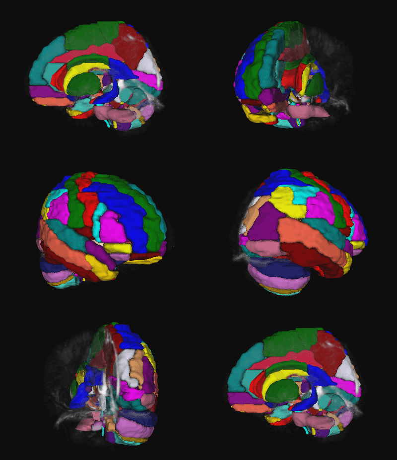
To this end, group-wise co-registered QSM templates were generated over various age intervals from age 1–83 years old. Registration was achieved by combining both T1-weighted and QSM images. Based on the proposed template, we created an accurate deep gray matter nuclei parcellation map (DGM map). Notably, we segmented thalamus into 5 sub-regions, i.e. the anterior nuclei, the median nuclei, the lateral nuclei, the pulvinar and the internal medullary lamina. Furthermore, we built a “whole brain QSM parcellation map” by combining existing cortical parcellation and white-matter atlases with the proposed DGM map.


Results:

Based on the proposed QSM atlas, the segmentation accuracy of iron-rich nuclei using QSM is significantly improved, especially for children and adolescent subjects. The age-related progression of magnetic susceptibility in each of the deep gray matter nuclei, the hippocampus, and the amygdala was estimated. Our automated atlas-based analysis provided a systematic confirmation of previous findings on susceptibility progression with age resulting from manual ROI drawings in deep gray matter nuclei. The susceptibility development in the hippocampus and the amygdala follow an iron accumulation model; while in the thalamus sub-regions, the susceptibility development exhibits a variety of trends.


Conclusion:

The common susceptibility atlases provided a standard coordinate system to conduct group analyses for QSM studies over a large age range. These atlases also provided an efficient tool for segmenting brain structures at specific ages, benefiting from the dramatic delineation between different brain tissues. The susceptibility atlases also indicated common tissue structure variations at each age interval, which is a critical reference for investigating the brain gray and white matter development with aging. A novel segmentation of the thalamus is provided.


Authors:

Yuyao Zhang 1, Hongjiang Wei 1, Matthew J. Cronin, Naying He, Fuhua Yan, Chunlei Liu *


Back to top

Imaging whole-brain cytoarchitecture of mouse with MRI-based quantitative susceptibility mapping.

Download PDF

Purpose:

The proper microstructural arrangement of complex neural structures is essential for establishing the functional circuitry of the brain. We present an MRI method to resolve tissue microstructure and infer brain cytoarchitecture by mapping the magnetic susceptibility in the brain at high resolution.


Methods:

In this study, mouse brains (n = 2) were scanned ex vivo at a nominal 10-μm isotropic resolution using a three-dimensional (3D) GRE sequence at 9.4 T. We applied STAR-QSM to address current issues of streaking artifacts.  In this dataset, QSM offers a powerful tool to resolve fine detailed magnetic susceptibility contrast in many structures, e.g. retina cell layers, olfactory sensory neurons, corpus callosum, putamen axon, cerebral cortical layers, barrel cortex, hippocampus layers, cerebellum, striatal neurons, and the brainstem. Using STAR-QSM, we are able to achieve in susceptibility mapping at a resolution and contrast exceeding traditional MR images.


Results:

We demonstrate magnetic susceptibility maps at a nominal resolution of 10-μm isotropic, approaching the average cell size of a mouse brain. The maps reveal many detailed structures including the retina cell layers, olfactory sensory neurons, barrel cortex, cortical layers, axonal fibers in white and gray matter. Olfactory glomerulus density is calculated and structural connectivity is traced in the optic nerve, striatal neurons, and brainstem nerves. The method is robust and can be readily applied on MRI scanners at or above 7 T.


Conclusion:

In this study, we demonstrate the utility of QSM in visualizing the microstructure of the intact mouse brain at a 10-μm resolution. QSM at near cellular resolution provides an exquisite delineation of brain microstructure, which overcomes limitations of current imaging methodologies. QSM offers a tool to assess the brain cytoarchitecture and the dataset achieved can serve as a reference for quantitative analysis of mouse brain microstructure.


Keywords:

Quantitative susceptibility mapping; Cytoarchitecuture; Mouse brain


Authors:

Hongjiang Wei, Luke Xie, Russell Dibb, Wei Li, Kyle Decker, Yuyao Zhang, G. Allan Johnson, Chunlei Liu *


Back to top

Susceptibility Tensor imaging and Tractography of Collagen Fibrils in the Articular Cartilage.

Download PDF

Purpose:

To investigate the B0 orientation-dependent magnetic susceptibility of collagen fibrils within the articular cartilage and to determine whether susceptibility tensor imaging (STI) can detect the 3D collagen network within cartilage.


Methods:

Multiecho gradient echo datasets (100-mm isotropic resolution) were acquired from fixed porcine articular cartilage specimens at 9.4 T. The susceptibility tensor was calculated using phase images acquired at 12 or 15 different orientations relative to B0. The susceptibility anisotropy of the collagen fibril was quantified and diffusion tensor imaging (DTI) was compared against STI. 3D tractography was performed to visualize and track the collagen fibrils with DTI and STI.


Results:

STI experiments showed the distinct and significant anisotropic magnetic susceptibility of collagen fibrils within the articular cartilage. STI can be used to measure and quantify susceptibility anisotropy maps. Furthermore, STI provides orientation information of the underlying collagen network via 3D tractography.


Conclusion:

The findings of this study demonstrate that STI can characterize the orientation variation of collagen fibrils where diffusion anisotropy fails. We believe that STI could serve as a sensitive and noninvasive marker to study the collagen fibrils microstructure.


Keywords:

diffusion tensor imaging (DTI); susceptibility tensor imaging (STI); magnetic susceptibility; magnetic susceptibility anisotropy; knee; collagen fibril; articular cartilage


Authors:

Hongjiang Wei, Eric Gibbs, Peida Zhao, Nian Wang, Gary P. Cofer, Yuyao Zhang, G. Allan Johnson, and Chunlei Liu *


Back to top

Precise Targeting of the Globus Pallidus Internus with Quantitative Susceptibility Mapping for Deep Brain Stimulation Surgery.

Download PDF

Objective:

The goal of this study was to demonstrate the use of quantitative susceptibility mapping (QSM)–based images to precisely localize the globus pallidus internus (GPi) for deep brain stimulation (DBS) planning and to enhance postsurgical visualization of the DBS lead positions.


Methods:

Presurgical T1-weighted (T1w), T2-weighted (T2w), and QSM images as well as postsurgical CT images were obtained in 29 patients with Parkinson’s disease. To enhance the contrast within the GP, a hybrid contrast was created by linearly combining T1w and QSM images. Contrast-to-noise ratios (CNRs) of the GPi on T1w, T2w, QSM, and hybrid images were compared. The CNR differences were tested using the 1-way ANOVA method. The visualization of the DBS lead position was demonstrated by merging the postsurgical CT with presurgical MR images.


Results:

The hybrid images yield the best CNRs for GPi depiction and the visualization of the postsurgical DBS lead position was significantly improved.


Conclusion:

QSM-based images allow for confident localization of borders of the GPi that is superior to T1w and T2w images. High-contrast hybrid images can be used for precisely directed DBS targeting, e.g., GPi DBS for the treatment of advanced Parkinson’s disease.


Keywords:

deep brain stimulation; quantitative susceptibility mapping; globus pallidus internus; Parkinson’s disease; electrode placement; functional neurosurgery; diagnostic technique


Authors:

Hongjiang Wei, Chencheng Zhang, Tao Wang, Naying He, Dianyou Li, Yuyao Zhang, Chunlei Liu, Fuhua Yan, and Bomin Sun


Back to top

Real-Time MRI reconstruction

Purpose:

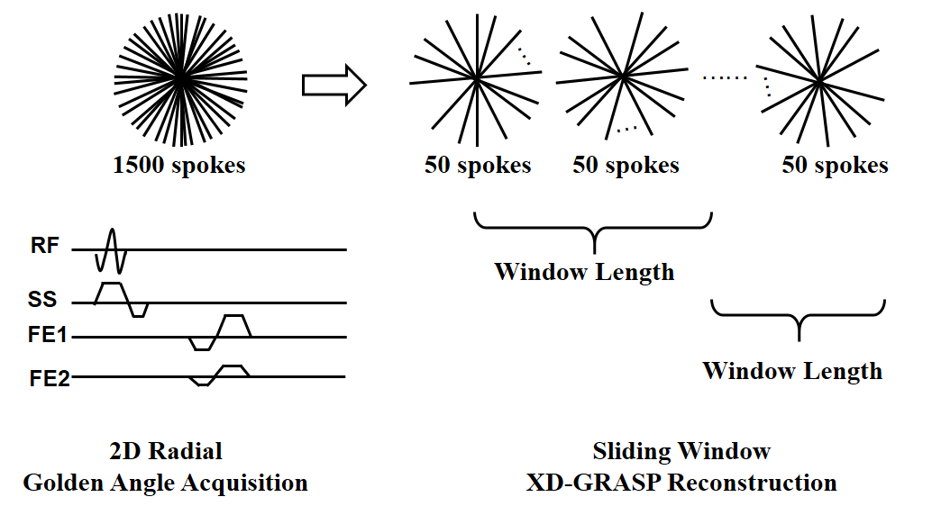
With the help of radial UTE sequence, the acquisition of MRI can be fast enough for real-time imaging. However, the image quality of radial UTE is unacceptable without suitable reconstruction method and the traditional reconstruction method cannot reach the request of real-time imaging. We try to develop a method to reconstruct high quality radial UTE MR images real-timely, which can serve as the key role of real-time invasive guidance.


Back to top